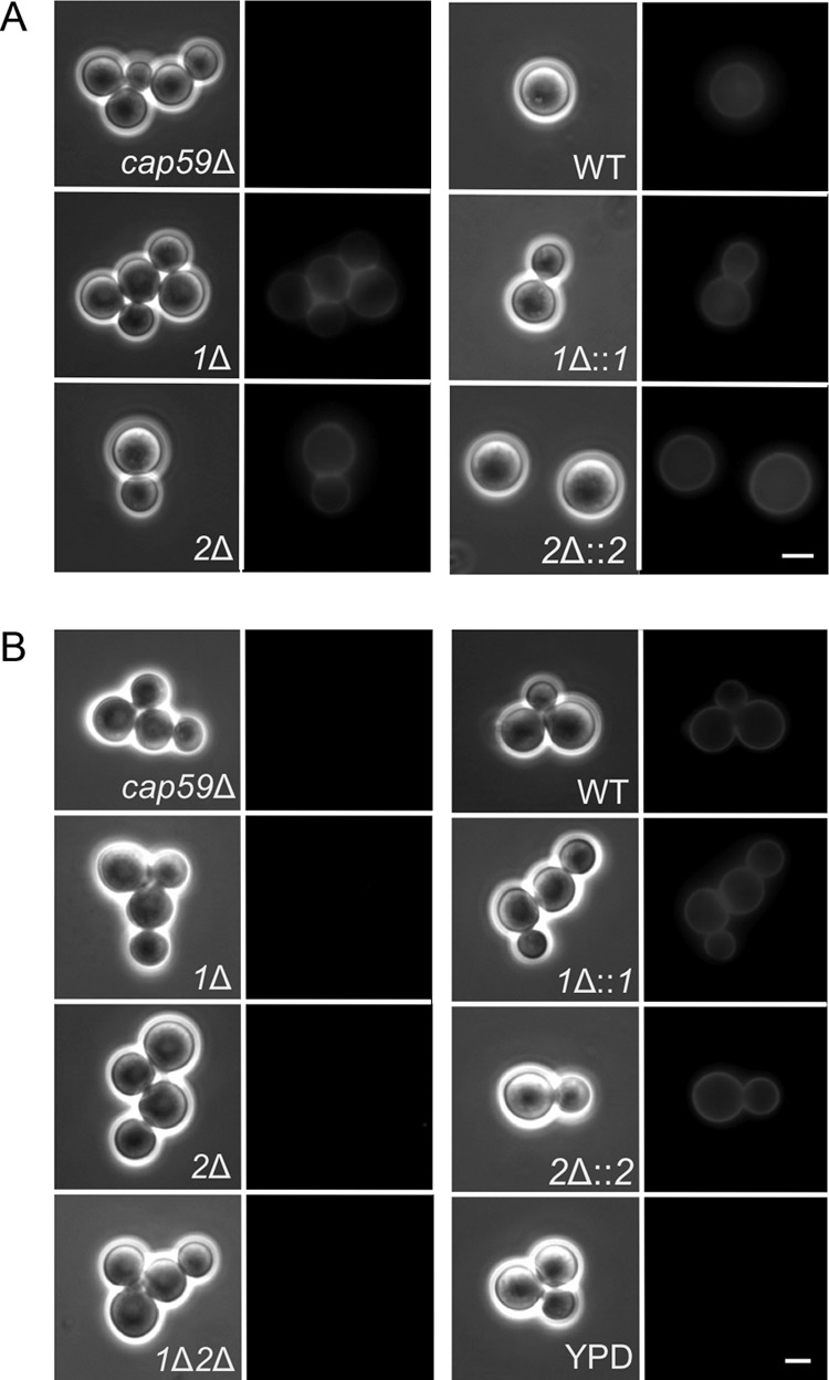
FIG 2.

pbxΔ strains display cell surface capsule but cannot donate capsule to acapsular acceptor cells in standard assays. (A) Surface capsule. Cells of the indicated strains were labeled with antibody 3C2 and imaged as described in the legend to Fig. 1. (B) Capsule transfer. CM from the indicated strains (or YPD alone as a control) was used as the capsule donor in transfer assays, with the cap59Δ mutant as the acceptor strain. Imaging is as described in the legend to Fig. 1. 1Δ, pbx1Δ mutant; 2Δ, pbx2Δ mutant; 1Δ::1, pbx1Δ mutant complemented with PBX1; 2Δ::2, pbx2Δ mutant complemented with PBX2; 1Δ2Δ, pbx1Δ pbx2Δ mutant. All images in both panels are at the same magnification. Bar = 5 μm.
