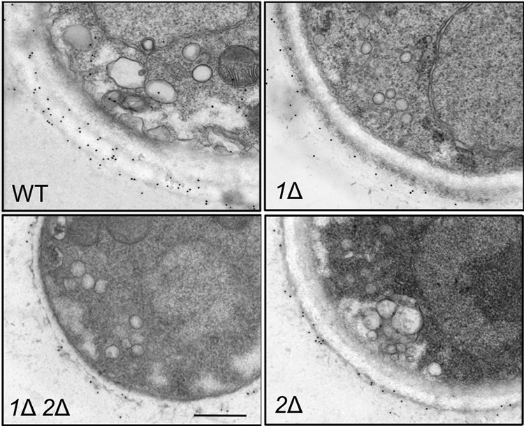
FIG 5.

pbxΔ mutants show reduced intracellular and surface capsule polysaccharide. Cryptococcal cells grown in YPD medium were examined by immunoelectron microscopy using colloidal gold-labeled anti-GXM monoclonal antibody 2H1. Each panel shows a portion of a cell of the indicated strain. All images are at the same scale. Bar = 500 nm.
