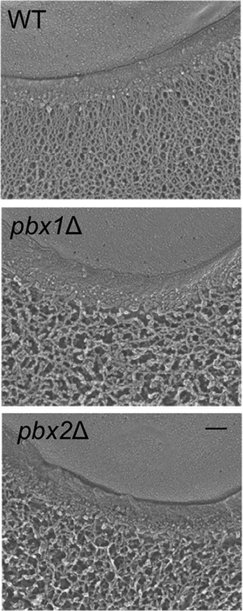
FIG 6.

The capsules of pbxΔ mutants have reduced density. Quick-freeze, deep-etch electron micrographs for the indicated strains are shown. Each image shows a portion of the cell edge, with the plasma membrane at the top and capsule fibers at the bottom (extending downwards) separated by the cell wall. All strains were grown under the same conditions, and all images are at the same magnification. Bar = 100 nm.
