FIG 8.

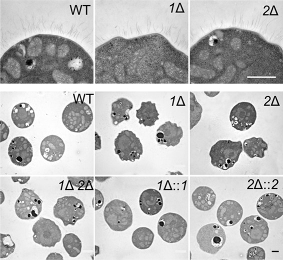
pbxΔ mutants show irregular cell morphology. Thin-section electron micrographs in the top row are all at the same magnification, and images in the bottom two rows are all at the same (lower) magnification. Bars = 1 μm.

pbxΔ mutants show irregular cell morphology. Thin-section electron micrographs in the top row are all at the same magnification, and images in the bottom two rows are all at the same (lower) magnification. Bars = 1 μm.