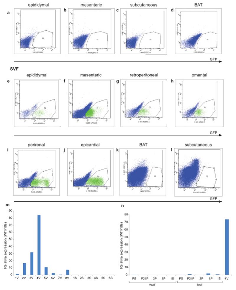
Figure 1. Wt1+ cells reside in the SVF in visceral WAT but not subcutaneous WAT or BAT depots.
Using the Wt1-GFP mouse model, representative FACS plots showing Wt1+ cells (GFP) are not detected in the mature adipocytes in the (a) epididymal, (b) mesenteric, (c) subcutaneous, or (d) BAT fat pad. In the SVF, Wt1+ cells are found in the (e) epididymal, (f) mesenteric, (g) retroperitoneal, (h) omental, (i) perirenal, and (j) epicardial depots; however, Wt1+ cells are not detected in the SVF from (k) BAT or (l) subcutaneous fat pads. GFP signal is indicated in the x-axis. Gates are chosen using cells from Wt1-GFP negative mice (n=3). (m) The level of WT1 mRNA in human visceral and subcutaneous fat is measured by Q-PCR (y-axis is expressed in arbitrary unit). V indicates ‘visceral’ (omental fat in this case). S indicates subcutaneous fat. (n), The level of WT1 mRNA from human ‘BAT’ and adjacent white adipose tissue (WAT) is measured by Q-PCR. Sample ‘4V’ is visceral fat which acts as a positive control (y-axis is expressed in arbitrary units).

