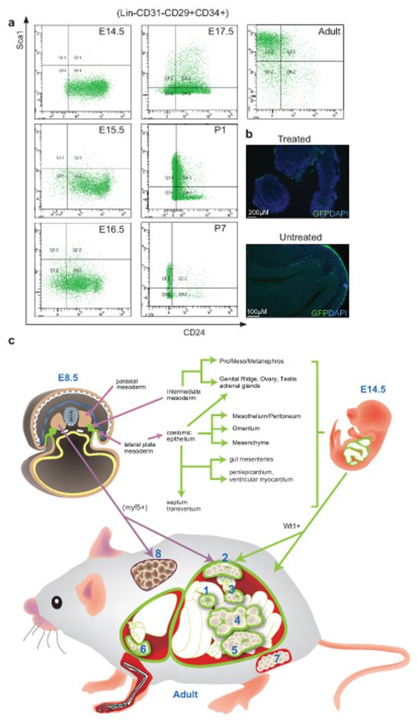
Figure 5. FACS profiling of mesothelium and mesothelium-derived cells by adipose progenitor markers.
(a) Mesothelium and the mesothelium-derived cells collected from the Wt1-GFP line at different developmental stages and adult were stained with adipose progenitor surface markers. Representative FACS plots showing GFP+ cells stained with the markers. (b) Immunofluorescence staining showing the mesothelium staining at the surface of visceral organs (E15.5), with and without collagenase treatment. GFP is indicated in green and cell nuclei are stained with DAPI (blue). (c) Summary of Wt1-expressing WATs and their possible origins during development. In E8.5, Wt1 is first expressed in the intermediate mesoderm and lateral plate mesoderm (highlighted in green). Wt1+ mesenchymal cells from intermediate mesoderm later give rise to pro/meso/metanephros, genital ridge, ovary, testis, and adrenal glands. Wt1-expressing coelomic epithelium (derived from lateral plate mesoderm) contributes to mesothelium, mesenchyme, and also genital ridge, ovary, testis, and adrenal glands. At E9.5, Wt1 expression is also found in septum transversum, which contributes to gut mesenteries, hepatic sinusoids, peri/epicardium, ventricular myocardium see note above, and diaphragm. At E14.5, Wt1 is expressed in the developing kidney, gonads, adrenal glands, spleen, omentum, mesenteries, the mesothelial layer of all visceral organs (including epicardium and pericardium), and the peritoneum, as well as mesenchyme located near these tissues; the mesothelium and mesenchyme are represented in green. In the adult, we found Wt1 expression in all the visceral fat pads including omentum (1), retroperitoneal (2), perirenal (3), mesenteric (4), epididymal (5) in the abdominal cavity, and epicardial (6) in the thoracic cavity. Its expression is not detected in the subcutaneous (7) and BAT (8) which are both outside the body cavity. It has been shown previously that the myf5+ population of cells in the paraxial mesoderm contributes to BAT as well as retroperitoneal WAT26. The origin of subcutaneous WAT (7) is currently not known.

