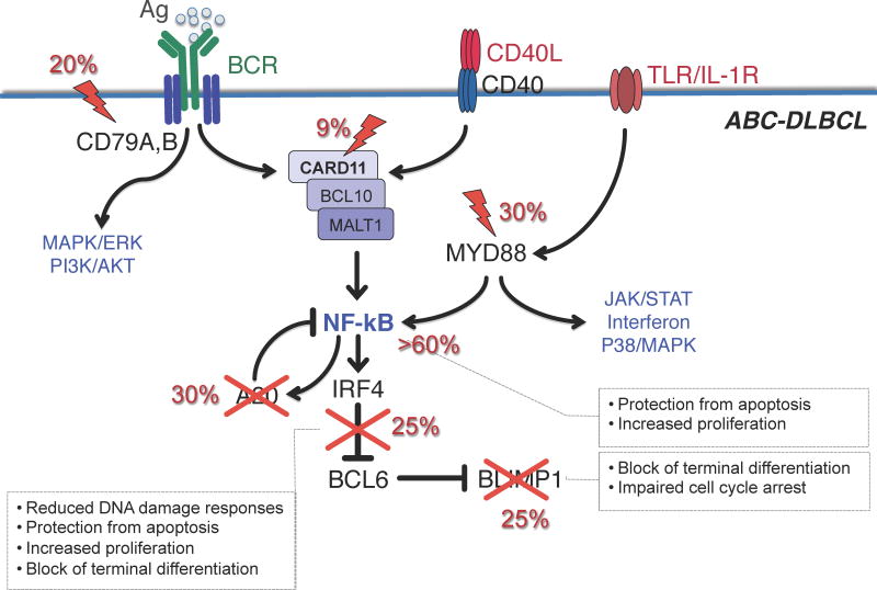
Figure 3. Pathway lesions in ABC-DLBCL.
Schematic representation of a GC centrocyte, expressing a functional surface BCR, CD40 receptor and TLR/IL-1R. Engagement of these signaling cascades in normal B cells converge on the activation of the NF-κB transcription complex, and induces the expression of hundreds of targets genes, including IRF4 and the NF-κB negative regulator TNFAIP3/A20. IRF4, in turn, downregulates BCL6 expression, allowing the release of the plasma cell master regulator PRDM1 and the development into a differentiated plasma cell. In DLBCL, a variety of genetic lesions disrupt this circuit at multiple levels specifically in the ABC-subtype, and contribute to lymphomagenesis by favoring the anti-apoptotic function of NF-κB while blocking terminal B cell differentiation. Crosses indicate inactivating mutations/deletions; lightning bolts denote activating mutations. Modified with permission from [12]

