Figure 6.

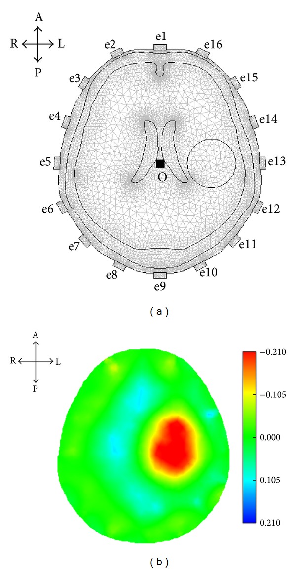
SEIT imaging based on simulated EIT data. (a) A simulated stroke lesion was set on the left side of the model. (b) The reconstructed SEIT image reflected the simulated lesion (A: anterior; P: posterior; L: left; and R: right).

SEIT imaging based on simulated EIT data. (a) A simulated stroke lesion was set on the left side of the model. (b) The reconstructed SEIT image reflected the simulated lesion (A: anterior; P: posterior; L: left; and R: right).