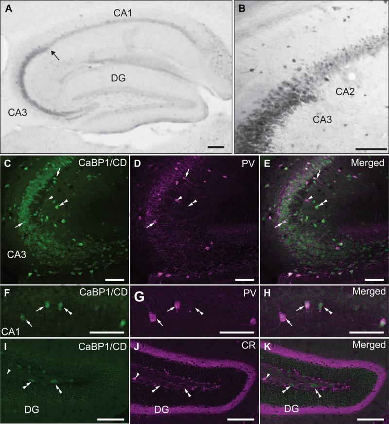
Figure 6. Localization of CaBP1/CD-IR in the hippocampal formation.
(A,B) Immunoperoxidase labeling with CaBP1/CD antibodies. Arrow in (A) indicates boundary between CA3 and CA2 regions, magnified in (B). (C-H) Double immunofluorescence labeling with CaBP1/CD antibodies (C,F) and parvalbumin antibody (PV; D,G) in CA3 (C-E) and CA1 regions (F-H). (I-K) Double immunofluorescence labeling with CaBP1/CD antibodies (I) and calretinin antibody (CR; J) in the dentate gyrus (DG). Arrows indicate neurons double-labeled for CaBP1/CD-IR and PV. Arrowheads indicate PV-positive (C-E) or CR-positive (I-K) neurons that do not show CaBP1/CD-IR. Double arrowheads indicate CaBP1/CD-positive neurons that are PV-negative (C-H) or CR (I-K). Scale bars, 100 μm.

