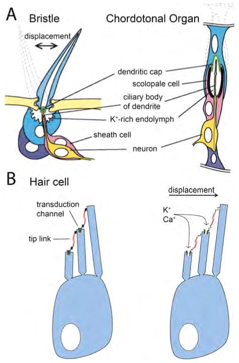
Fig. 2.
Mechanosensory cells. A) A depiction of two mechanosensory organs in D. melanogaster, and their proposed mechanisms of activation upon position change (shown by dotted outlines) as a result of a mechanical stimulus. In both organs, the neuronal dendrite is attached to a dendritic cap that is affected by stimulus-induced movement, activating mechanotransduction channels. Adapted with permission from: Jarmen A. N. et al. Human Molec. Genetics 2002; 11:1215–1218. B) A vertebrate hair cell before (left) and after (right) displacement by endolymph movement. The shear force pushing stereocilia creates tension in a spring-like element (as yet unidentified) that is proposed to be in series with the tip link. Increased tension results in transduction-channel opening.

