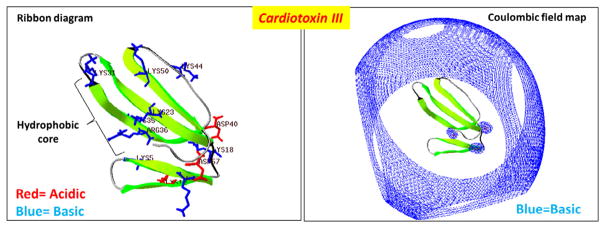
Figure 1. A rendering of the 3D structure of cardiotoxin III from Naja naja atra.

A ribbon diagram of the crystal structure of cardiotoxin III (PDB #2CRT) is illustrated to highlight the hydrophobic core region and show basic residues in blue and acidic residues in red. Right panel: A rendering of the Coloumbic electrostatic field potential map for cardiotoxin III which shows that the toxin possesses a very extensive and wide basic electrostatic field potential landscape (blue). Electrostatic field potential calculations, 3D molecular rendering, and specific annotations of cardiotoxin III was performed by using the Swiss PDB Viewer software.
