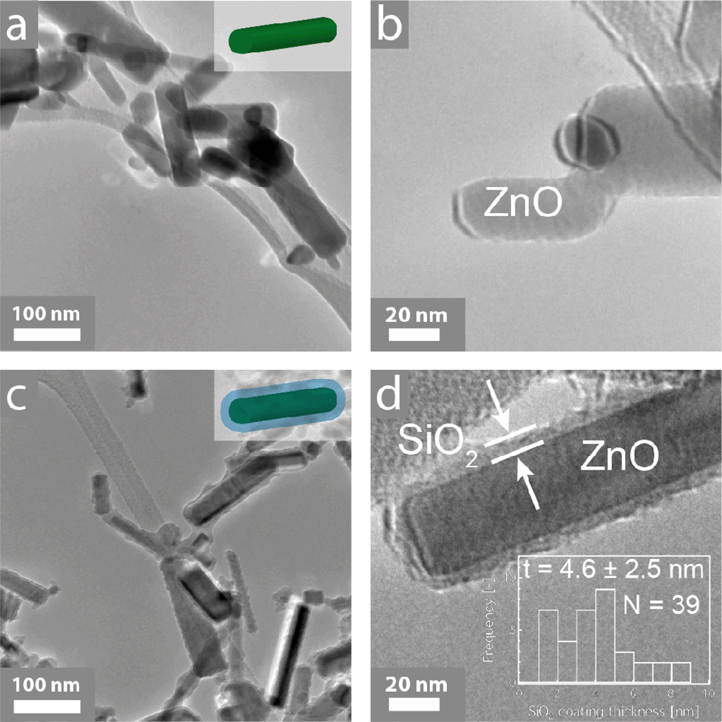
Figure 1.
Transmission electron microscopy images of the uncoated (a, b) and SiO2-coated ZnO nanoparticles. In both cases, the ZnO forms nanorods with aspect ratio ~3:1. Furthermore, there is a nanothin amorphous SiO2 shell encapsulating the core particles for the SiO2-coated ZnO nanorods. In the inset of (d) the SiO2 coating thickness distribution along with the average coating thickness ± standard deviation and the total number N of coatings counted.

