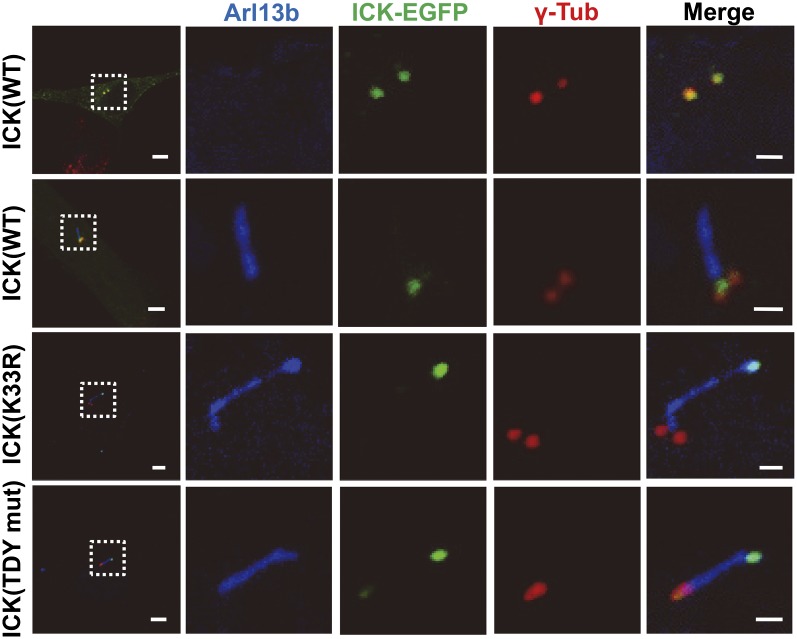
Fig. 3.
ICK activity is required for its ciliary localization. To determine the subcellular localization of WT or mutant (K33R or TDY mut) ICK in primary cilia, cultured cells were transfected with appropriate plasmids and immunostained with Arl13b (blue) and γ-tubulin (γ-Tub, red) antibodies for cilia and basal bodies, respectively. Wild-type ICK was localized near the basal body of shortened cilia. By contrast, inactive ICK mutants were localized at the distal tip of cilia, which displayed swollen morphology. (Scale bars: 5 µm, Left; 1 µm, Right.)

