Fig. 3.
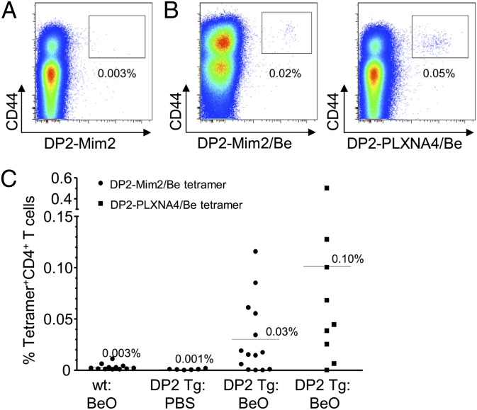
Be-responsive CD4+ T cells in the lung of HLA-DP2 transgenic (Tg) mice recognize the same αβTCR ligand as their counterparts in human lung. (A) Representative density plots depicting ex vivo lung CD4+CD44hi T cells from a BeO-exposed HLA-DP2 Tg mouse stained with an HLA-DP2–mimotope-2 tetramer without loaded Be. (B) Representative density plots of ex vivo lung cells from a BeO-exposed HLA-DP2 Tg mouse stained with either a Be-loaded HLA-DP2–mimotope-2 (DP2-Mim2/Be; Left) or HLA-DP2–plexin A4 (DP2-PLXN4/Be; Right) tetramer. (C) Cumulative percentage of ex vivo CD4+ T cells from the lungs of PBS- or BeO-treated WT FVB/N and HLA-DP2 Tg mice positively staining with HLA-DP2/mimotope-2/Be and HLA-DP2–PLXNA4/Be tetramers is shown.

