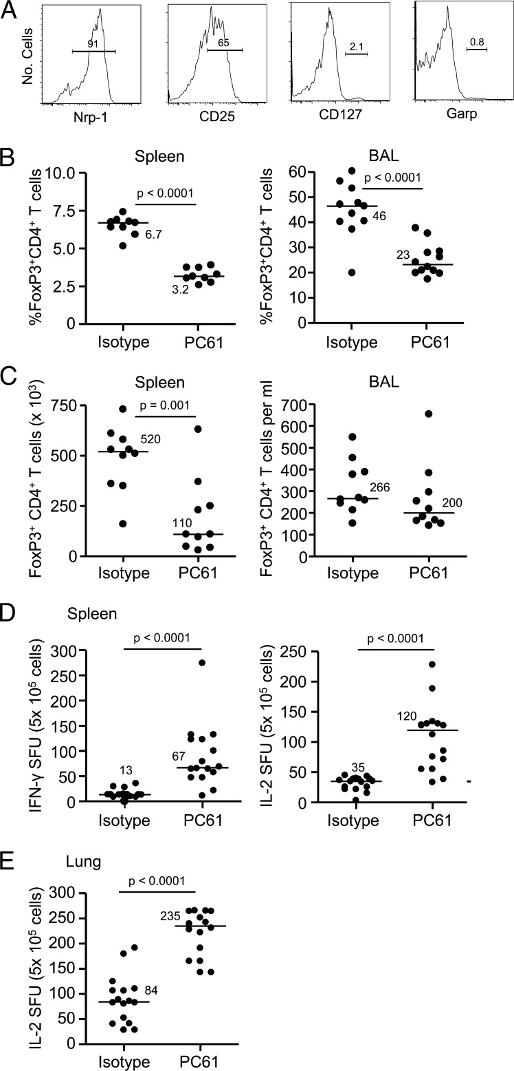
Fig. 4.
Treg cells are expanded in the lung of BeO-exposed HLA-DP2 transgenic (Tg) mice and modulate Be-specific CD4+ T cells. (A) Representative histograms of neuropilin-1 (Nrp-1), CD25, CD127, and Garp expression on FoxP3+ CD4+ T cells in the BAL of BeO-exposed HLA-DP2 Tg mice are shown. (B) Percentage of FoxP3+ CD4+ T cells in the spleen (Left) and BAL (Right) at day 21 of BeO exposure in HLA-DP2 Tg mice after exposure to isotype control or PC61 is shown. (C) Number of FoxP3+ CD4+ T cells in the spleen (Left) and BAL (Right) at day 21 of BeO exposure in HLA-DP2 Tg mice after exposure to isotype control or PC61 is shown. Frequency of Be-specific T cells in spleen (D) and lung (E) of BeO-exposed HLA-DP2 Tg mice using IFN-γ and IL-2 ELISPOT 21 d after exposure to isotype control or PC61 is shown. Data are expressed as the mean spot-forming units (SFUs) per 5 × 10⁵ cells. For B–E, median values are indicated with solid lines, and data shown represent at least nine mice per group from two separate experiments.

