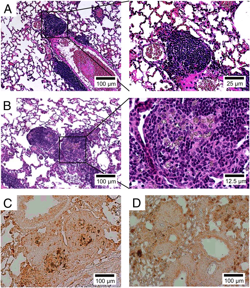
Fig. 6.
Treg cells modulate Be-induced granulomatous inflammation. Representative H&E staining of lung from BeO-exposed HLA-DP2 transgenic (Tg) mice that were treated with an isotype control (A) or an anti-CD25 (B) mAb is shown. Magnification (10×) is shown (A and B, Left) and an enlarged view (40× magnification) of the area enclosed within the black box (A and B, Right) is shown. Representative immunohistochemical staining for the macrophage marker, Mac-1, is shown in lung tissue obtained from BeO-exposed HLA-DP2 Tg mice with (C) and without (D) treatment with an anti-CD25 depleting mAb, PC61, at 21 d. Histology images are representative of a total of at least nine mice from each treatment group from three separate experiments.

