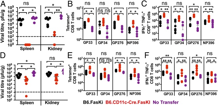
Fig. 5.
Transfer of Fas-negative DCs rapidly clears an established LCMV-clone 13 infection. B6 mice infected with LCMV-clone 13 for 8 d were injected with 2 × 10⁶ purified splenic DCs (A–C) or GM-CSF–derived BMDCs (D–F) from uninfected B6.FasKI or B6.CD11c-Cre.FasKi animals or received no transfer. (A and D) Recipient groups of B6 mice were analyzed 3 wk after DC transfer and viral titers were estimated in the indicated organs. Dotted line indicates the limit of detection. (B and E) LCMV-specific T cells were estimated in the spleens of recipient mice by staining with indicated tetramers. (C and F) IFN-γ and TNF-α double-producing T cells numbers were identified by intracellular flow cytometric analysis after 5 h in vitro stimulation with indicated LCMV peptides. Data are compiled from two independent experiments with 5–8 mice per time point. Error bars represent SEM. Statistical significance was determined by ordinary one-way ANOVA (*P < 0.05; **P < 0.005; ns, not significant).

