Fig. 2.
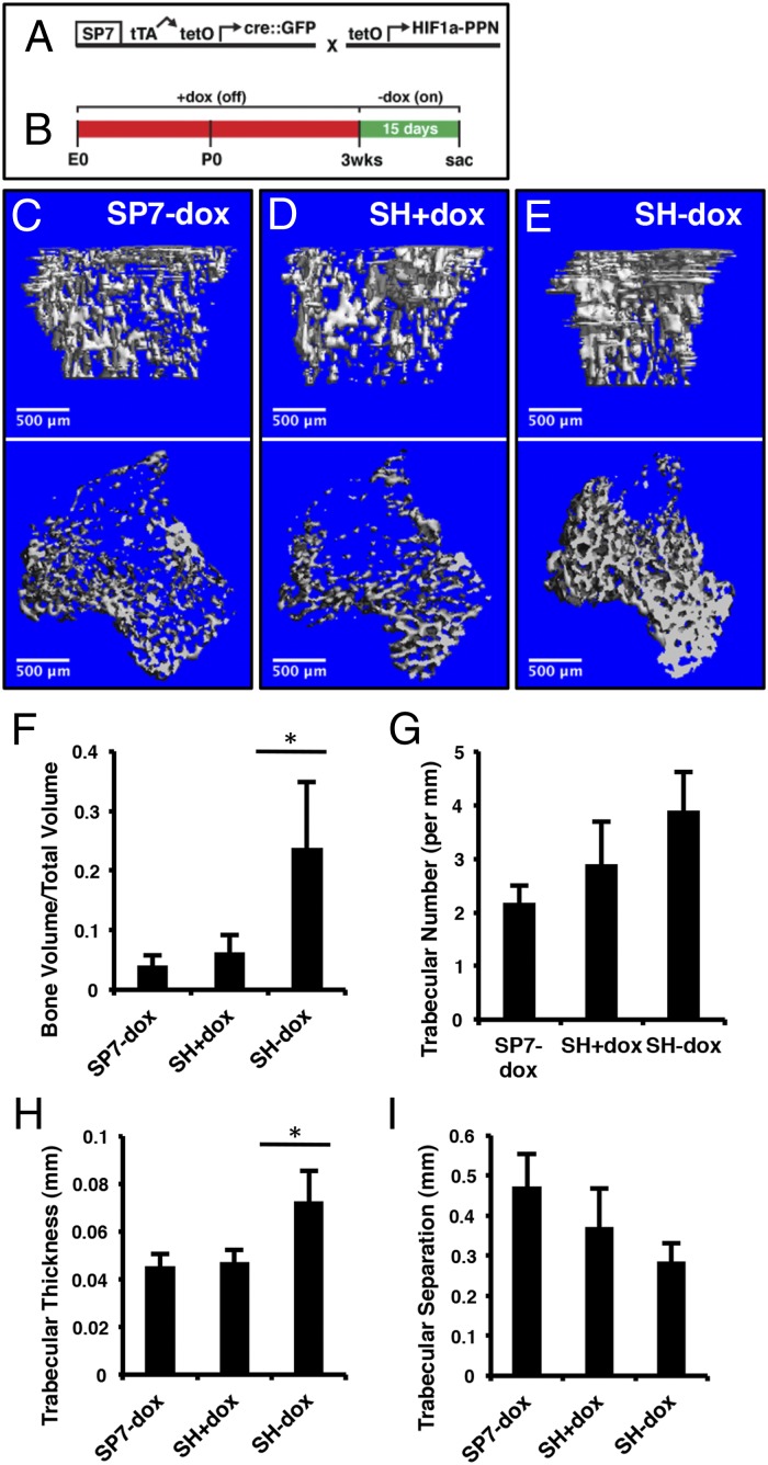
Expression of HIF1-PPN in SP7-positive cells increases cancellous bone volume. (A) Schematic of transgenes used to yield SP7;HIF1-PPN (SH) mice. The tetO-driven HIF1-PPN is only expressed in cells actively transcribing the SP7 promoter-driven tTA. (B) Treatment schematic. The experimental window began at 3 wk of age with dox removal. Mice were analyzed 15 d later. (C–E) µCT analyses of 1.6 mm of cancellous bone immediately under the growth plate of the tibia. Mouse genotypes are as labeled. In D, SH mice were maintained on dox throughout the experimental window to evaluate leakiness in the suppression system. (F) Bone volume per total volume of the regions shown in C–E. There was no difference between SP7-dox and SH+dox mice but a significant increase (P = 0.0015) in SH-dox animals. (G) Trabecular number per millimeter, as quantified from µCT scans. (H) Trabecular thickness by µCT (P = 0.0010). (I) Trabecular separation. n = 7 (SP7-dox); n = 6 (SH+dox); n = 6 (SP7-dox).

