Fig. 6.
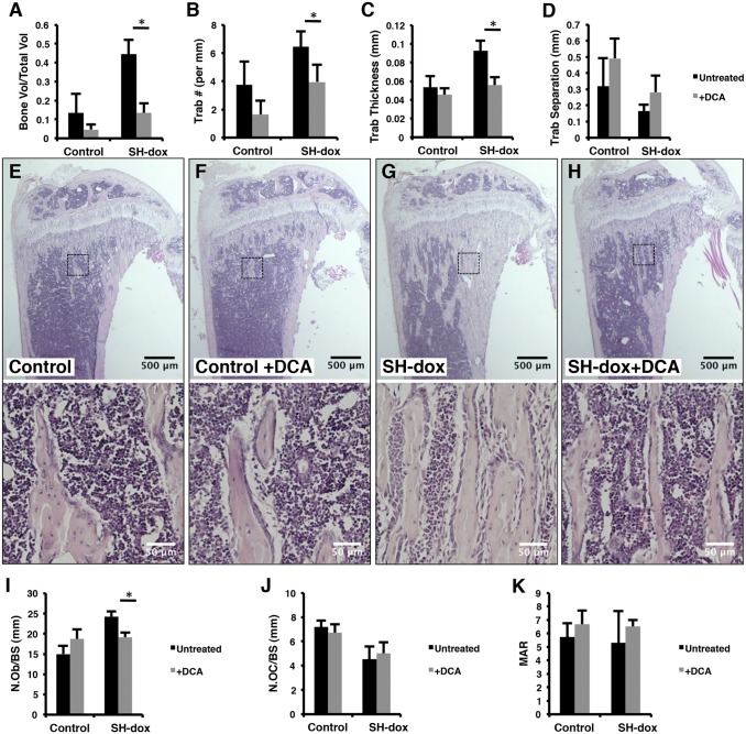
PDK1 inhibition blocks the bone anabolic effects of HIF1-PPN. (A–D) Quantification of µCT results. DCA treatment significantly reduced the BV/TV, trabecular number, and trabecular thickness in SH-dox mice (P = 0.0005, P = 0.0232, and P = 0.0018, respectively). BV/TV in control mice was also reduced with DCA treatment, but this number did not reach significance (P = 0.0921). n = 4 (control, SH-dox, SH-dox+DCA); n = 5 (control+DCA). (E–H) Histological sections of the tibia with higher magnification views of the boxed regions shown in the lower panels. (I) Osteoblast numbers per bone surface, as evaluated by histomorphometry. DCA treatment significantly lowered osteoblast numbers in SH-dox mice (P = 0.0327). (J) Osteoclast numbers per bone surface, as evaluated by histomorphometry. n = 3 (control, SH-dox+DCA) and n = 4 (control+DCA, SH-dox) for cell counting. (K) Mineral apposition rate (MAR) as determined by calcein double labeling. No significant changes were observed. Control mice were sex-matched littermates without SP7-tTA, subjected to same dox regime as experimental groups.

