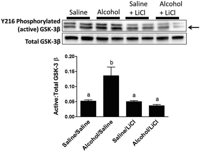
Fig. 7.
Western blot analysis of fracture callus active GSK 3β levels. Representative western blots for Tyr216-phosphorylated (activated) and total GSK-3β protein levels in fracture callus lysates at Day 9 post-fracture. Data are presented as the densitometric ratio of activated/total GSK-3β. Only the bottom band (arrow) was quantified, as cross-reactivity with the heavier GSK-α also occurs with this antibody. Alcohol exposure resulted in a significant increase in the ratio of activated GSK-3β compared with saline-exposed mice. LiCl treatment significantly decreased activated GSK-3β to levels found in saline-exposed mice. LiCl treatment did not change GSK-3β in the saline-exposed group. Groups not sharing a letter are significant, P ≤ 0.05 using one-way ANOVA and Tukey's multiple comparison procedure. N = 4–8 mice/treatment group.

