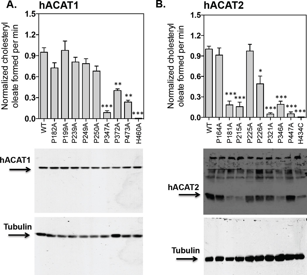
Figure 5. Protein expression and enzymatic activities of various hACAT1 or hACAT2 proline mutants within the TMDs.
The ACAT deficient mutant CHO cells (AC29) were transiently transfected with various hACAT1 plasmids (A) or various hACAT2 plasmids (B) as indicated. The procedures for transfection and for measuring ACAT enzyme activity in intact cells are described in Materials and Methods, as demonstrated in Fig. 2. By western analysis, hACAT1 is identified as a single band at 50 kDa; hACAT2 is identified as a single band at 46 kDa. The anti-ACAT1 antibodies used here were high-titer antibodies. Instead, the anti-ACAT2 antibodies used here were not high-titer antibodies; in addition to the ACAT2 protein band, these antibodies also detected several non-specific bands located at regions above the ACAT2 protein band. Values for the WT hACAT1 or WT hACAT2 activity were set 1.0. The western blot results are from one single experiment representative of two separate experiments. The enzyme activity data are reported as means ± SEM and are averages of two separate experiments. The p values were obtained by comparing values of individual mutant hACATs vs value of WT hACAT. ** P<0.01; *** P<0.001.

