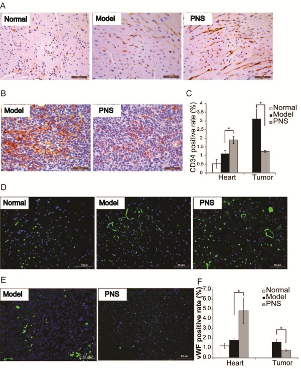
Figure 3.

The expression of CD34 and vWF in the heart and tumor in the complex mouse model. Immunohistochemistry examination of heart A. and tumor B. for the expression of CD34 was examined. CD34-positive cells appeared in brown. Nuclei were stained as blue by hematoxylin. C. Quantification of CD34 positive staining in the heart and tumor, respectively. Immunohistochemistry examination of heart D. and tumor E. for the expression of vWF was also examined. vWF -positive cells appeared in green. Nuclei were stained as blue by DAPI. F. Quantification of vWF positive staining in the heart and tumor, respectively. Scale bars indicate 50 μm. *p < 0.05, compared to the vehicle-treated complex model.
