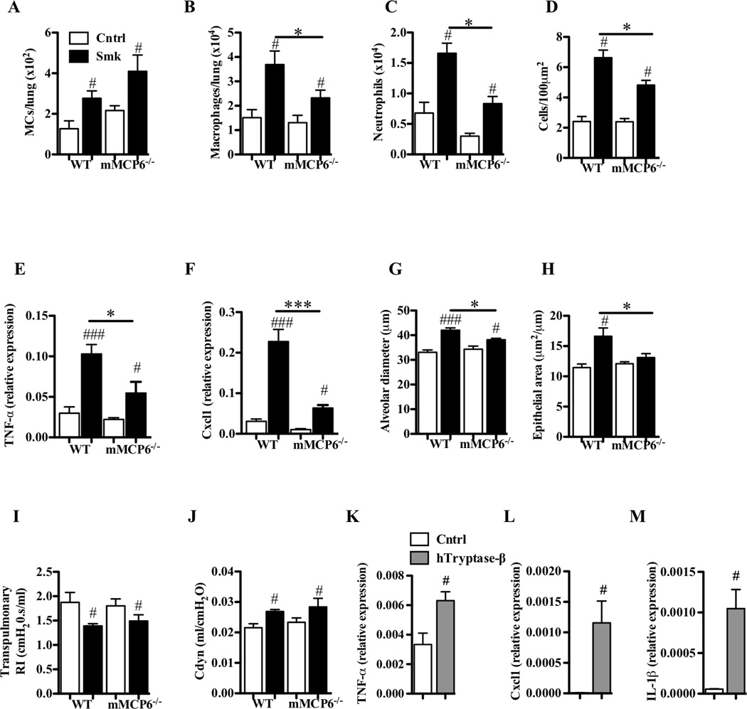
FIG 8.
The tryptase mMCP-6 contributes to pulmonary macrophage accumulation and parenchymal inflammation, and is required for airway remodeling and alveolar enlargement in experimental COPD. A–J, WT and mMCP-6−/− B6 mice were exposed to cigarette smoke or normal air for 8 weeks. Relative to smoke-exposed WT mice, smoke-exposed mMCP-6−/− mice had (A) no change in the number of Kit+/FcεRI+/IgE+ MCs but had reduced (B) numbers of F4/80+ macrophages and (C) neutrophils in the lung, (D) less cellular infiltrates in the parenchyma, attenuated (E) TNF-α and (F) Cxcl1 mRNA levels in lung homogenates, (G) diminished alveolar enlargement, (H) no airway remodelling and no differences in lung function [e.g. (I) transpulmonary resistance (RI) or (J) dynamic compliance (Cdyn)]. K–M, B6 mouse bone marrow-derived macrophages were cultured in the absence or presence of recombinant hTryptase-β. Relative to untreated cells, hTryptae-β-treated cells had increased levels of the transcripts that encode (K) TNF-α, (L) Cxcl1 and (M) IL-1β. Data are means±SEM of 6–8 mice/group, or of 3 cell cultures in triplicate (representative of 4 repeat experiments), # P<0.05, ## P<0.01, ### P<0.001 compared to mice that breathed normal air (A–J) or compared to sham-treated macrophages (K–M), * P<0.05, compared to other groups indicated.

