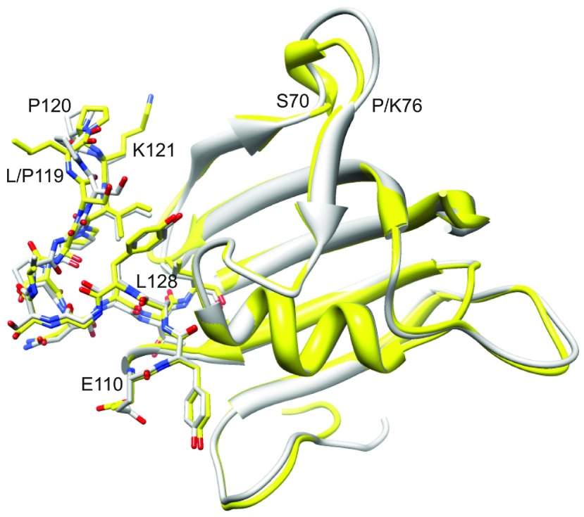
Figure 4. Superimposition of the FK1 domains of FKBP51 and FKBP52.
The FKBP51 X-ray structure from PDB code 3O5P [28] is illustrated in yellow, whereas molecule A from PDB code 4LAV [33] for FKBP52 is shown in grey. All heavy atoms are illustrated for the β4–β5 loop extending from Glu110 to Leu128. Substantial deviations in backbone geometry are only apparent for the β3 bulge (Ser70–Lys76) and the tip of the β4–β5 loop.

