
Figure 3.
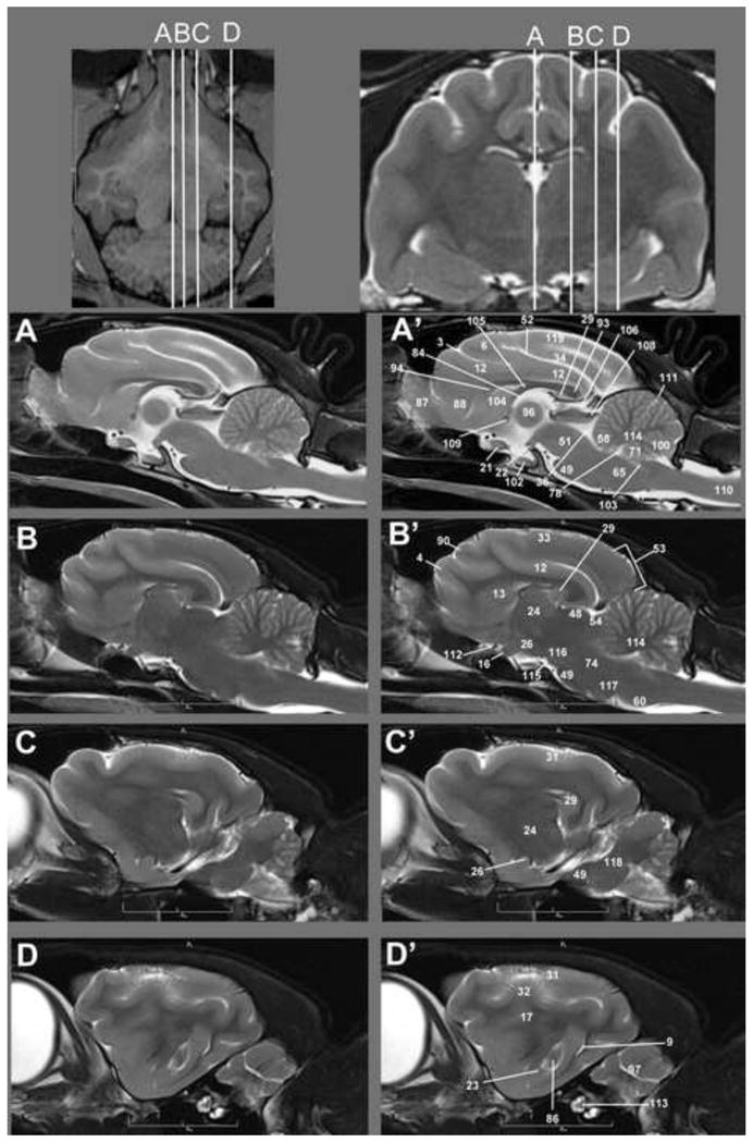
Sagittal T2w images of the cat brain. Top: Dorsal and transverse localizer denoting location of dorsal sections. Sections begin on midline (A) and extend out laterally through the hemisphere (D) Unlabeled images are located left (A–D) of the labeled MRI images (right A′-D′). Identified anatomical structures are listed in the image key and numbering is consistent throughout images.
3D Neuroimaging. 3D T1w MPRAGE MRI. Sagittal, transverse and dorsal planes are available to be visualized simultaneously. Crosshairs denotes area of brain examined and its location in all three views.