
Figure 1.
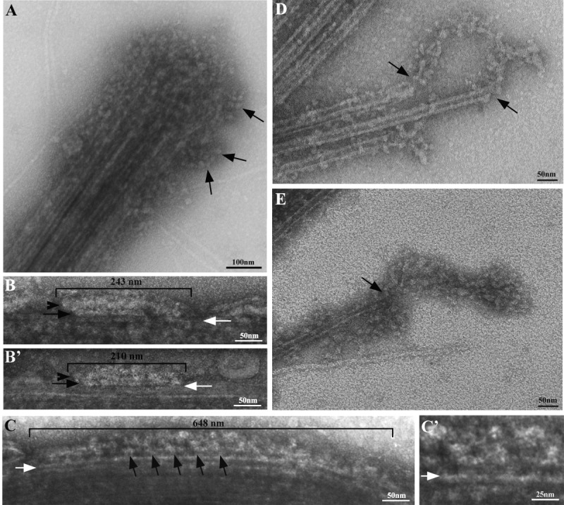
Negative staining of in situ IFT particles. A. After treatment with 0.1% glutaraldheyde and 0.1% NP-40, the axoneme tip is compact and still embedded in a mass of roundish particles; a few strings of particles project laterally from the tip (arrows). B–B', C. After demembranation in the presence of glutaraldheyde, linear arrays of particles are still associated with the side of the axoneme; their length and organization are compatible with those previously described for either short or long IFT trains [Pigino et al., 2009]. B–B'. Short arrays of particles are indicated by a bracket over which the length of the array is reported. The black arrows indicate the longitudinal domain present in the short arrays only, which is located between particles and the microtubule surface. Arrowheads indicate the thread of IFT particles located between the rod-like domain and membrane remnants. C. A long array of particles (bracket). The length of the array is indicated. Arrows point to the regular patterning of particles that is more clearly visible in the middle part of the array. The latter region is shown at a greater magnification in C'. White arrows in B–B', C–C' mark the microtubular surface. Images are oriented so that the tip of flagellum points to the left of the plate. D–E. At lower fixative and detergent concentrations (0.04% glutaraldehyde and 0.05% NP-40), the axoneme tip is partly disorganized and microtubule doublets splay onto the grid, showing strings of particles associated with the end of the tubule (arrows).