
Figure 4.
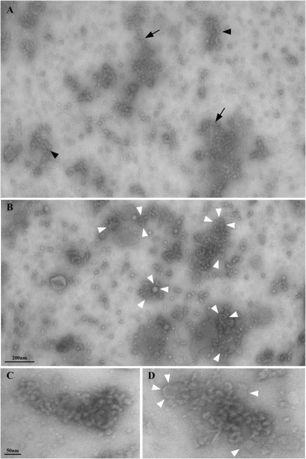
A–B. Low magnification electron microscopy views of the material in the gradient pellet after negative staining, showing the presence of sinuous arrays of particles with different lengths and spatial arrangements. Arrows in A point to single strings of particles, while arrowheads indicate double strings or thread-like arrays of particles. In B the frequent association of particles with membrane vesicles (white arrowheads) is shown. Higher magnification views of two arrays of particles are shown in C–D; white arrowheads in D indicate membrane vesicles.