
Fig. 4.
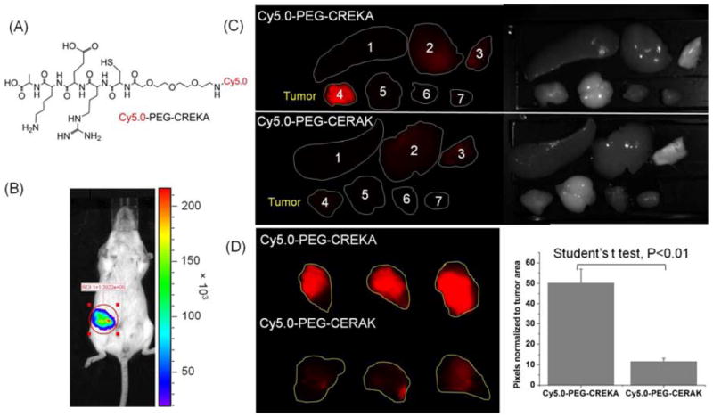
(A) The structure of Cy5.0-PEG-CREKA. (B) Bioluminescence images showing mice bearing 4T1-Luc breast tumor in mammary fat pad two weeks after implantation. Mice were intraperitoneally injected with D-luciferin (150 mg/kg body weight) 10 min before imaging with Xenogen IVIS Lumina system. (C) Mice bearing 4T1-GFP-Luc2 breast tumor were intravenously injected with Cy5.0-PEG-CREKA or Cy5.0-PEG-CERAK (0.3 μmol/kg body weight). After 4 h, the mice were sacrificed and the tumors and various organs (1. Spleen, 2. Liver, 3. Lung, 4. Tumor, 5. Brain, 6. Muscle, 7. Heart) were imaged with the Maestro FLEX In Vivo Imaging System. (D) Tumors of mice injected with Cy5.0-PEG-CREKA or Cy5.0-PEG-CERAK were imaged side by side with the Maestro FLEX In Vivo Imaging System and the pixels normalized to tumor area were analyzed with Maestro software (n = 3).