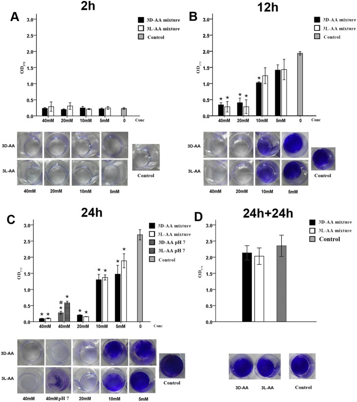
Figure 3. Effect of the mixture of D-Cys, D-Asp, and D-Glu at 40 mM (3D-AA) and the mixture of L-Cys, L-Asp, and L-Glu at 40 mM (3L-AA) on S. mutans biofilms.
S. mutans culture and BHIS at a ratio of 1∶100 were added to 96-well plates (semi-quantitative analysis) and a 24-well microplate (visualization) and were challenged with different concentrations of 3D-AAs or 3L-AAs for 2 h (A), 12 h (B), and 24 h (C), and the S. mutans biofilm formation was quantitatively evaluated by crystal violet staining. (D) Effect of 3D-AAs or 3L-AAs at 40 mM on existing S. mutans biofilms. S. mutans culture and BHIS at a ratio of 1∶100 were cultured for 24 h. The existing biofilm was exposed to 3D- or 3L-AAs for 24 h and quantified by CV staining. The results are an average of three independent experiments repeated five times, and mean and standard deviation were shown. “*” indicates a significant difference in the OD value of S. mutans biofilm between the groups challenged with 3D- or 3L-AAs and the control group, and a value of p<0.00625 was considered significant; “#” indicates a significant difference in the OD value of the S. mutans biofilm between the groups challenged with 3D-AA and 3L-AA at pH 7, and a value of p<0.05 was considered significant.

