
Figure 1. Clinical characteristics of two patients in Chinese CD family A.
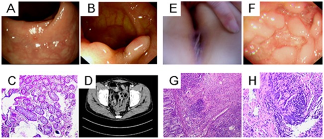
The index patient in the family, the father (Panels A to D) had evidence of mucous membrane granulation, polypoid proliferation and hyperemia in his colonoscopy, as shown in Panels A and B. Panel C shows the patient’s pathological findings of chronic intestinal inflammation. Panel D shows the thickening of the ileum wall by small intestine computed tomography enterography (CTE). The daughter in the family is another Patient (Panels E to H). Panel E shows her anal fistula at disease onset. Endoscopy showed intestinal poly-ulcers in Panel F. A biopsy showed non-specific granulomatous inflammation, as shown in Panel G, and the higher magnification of the pathology shown in Panel H reveals negative acid-fast staining granulomas. All of the images were collected in March 2012 in Zhongnan Hospital of Wuhan University.