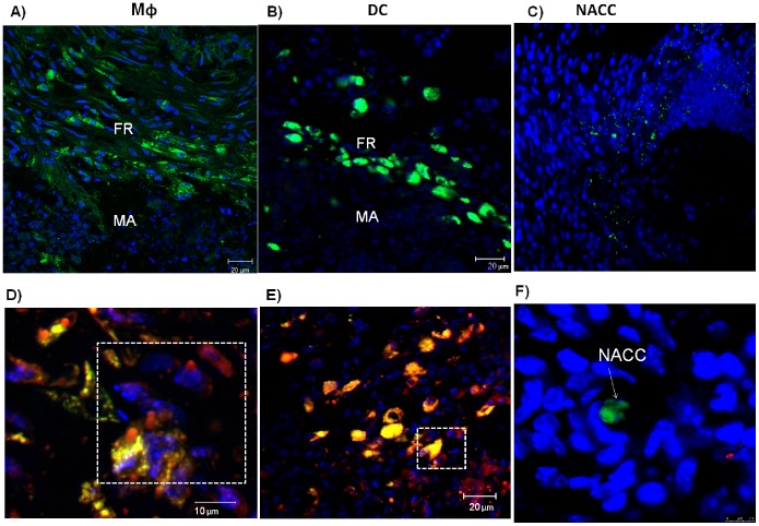
Figure 5. Transferred CFSE-labeled macrophages (Mφ) and dendritic cells (DC) become lipid-laden.
Macrophages (Mφ; in A) and dendritic cells (DC; in B) transferred at day 30 of a N. brasiliensis infection are 7 days later localized in the fibrotic ring (FR) of a microabscess (MA). Non-adherent spleen control cells (NACC; in C) transferred at day 30 of a N. brasiliensis infection are 7 days later localized outside of the FR and MA. Nile Red staining of lipid droplets is observed in transferred Mφ (D) and DC (E). Transferred cells are stained green by CFSE, lipid droplets are stained red by Nile Red, and nuclei are stained blue with DAPI.

