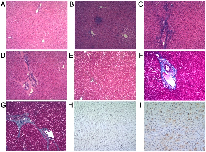
Figure 2. Liver histology. A–D (H & E stain, original magnification, ×10), E–G (Masson’s trichrome stain, original magnification, ×10), H–I (Immunohistochemistry, original magnification, ×40).
(A) Liver section from a control rabbit with no visible pathological signs of HEV infection. (B)-(C) Lymphocytes distributed focal or scattered in hepatic lobule, the inflammatory cells gathered along blood vessel walls. (D) Chronic inflammatory cells infiltrate the portal area, blood vessel walls thickening associated with fibrosis, local hyaline degeneration. (E) No histopathological changes with minimal staining limited to areas immediately adjacent to portal structures. (F) Artery wall thickening associated with moderate to severe fibrosis. (G) More advanced portal and periportal fibrosis with short fibrous septa. (H) Negative immunohistochemistry result for HEV antigen in liver sections from the control rabbits. (I) Positive results for HEV antigen in liver sections of experimental groups.

