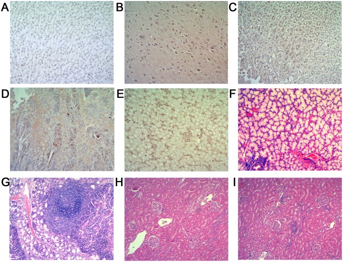
Figure 3. Extrahepatic tissue histology. A–E (Immunohistochemistry, original magnification, ×40), F–J (H & E stain, original magnification, ×10).
(A) Negative immunohistochemistry result for HEV antigen in extrahepatic tissue sections from the control rabbits. (B)–(E) Positive results for HEV antigen in brain, stomach, duodenum and kidney. (F) Duodenum section from a control rabbit with no visible pathological signs of inflammation. (G) A large number of lymphocytes infiltrate mucosal interstitial, focal lymph follicles formed in duodenum sections. (H) Kidney section from a control rabbit with no visible pathological signs of HEV infection. (I) Multifocal lymphocytes and mononuclear cells infiltrate in renal interstitial.

