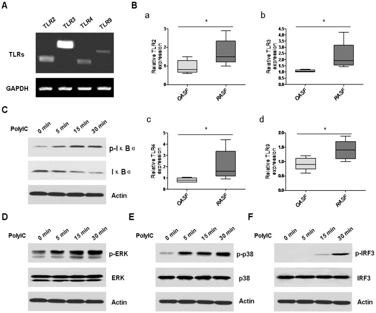
Figure 1. Functional TLRs were excessively expressed in RASF.
A, The expression of TLRs, including TLR2, TLR3, TLR4, and TLR9 in RASF was detected by RT-PCR analysis. GAPDH was used as the loading control. B, The expression of TLR2, TLR3, TLR4, and TLR9 in RASF (n = 5) and OASF (n = 5) was determined by realtime PCR (Wilcoxon signed-rank test, *P<0.05). RASF were stimulated with 25 µg/ml ploy(I:C) for 5 min, 15 min, and 30 min, respectively. Then the cells were harvested and lysised for western blot analysis of p-IκBα (C), p-ERK (D), p-p38 (E), and p-IRF3 (F). β-actin was used as the loading control. The results were representative of at least three experiments.

