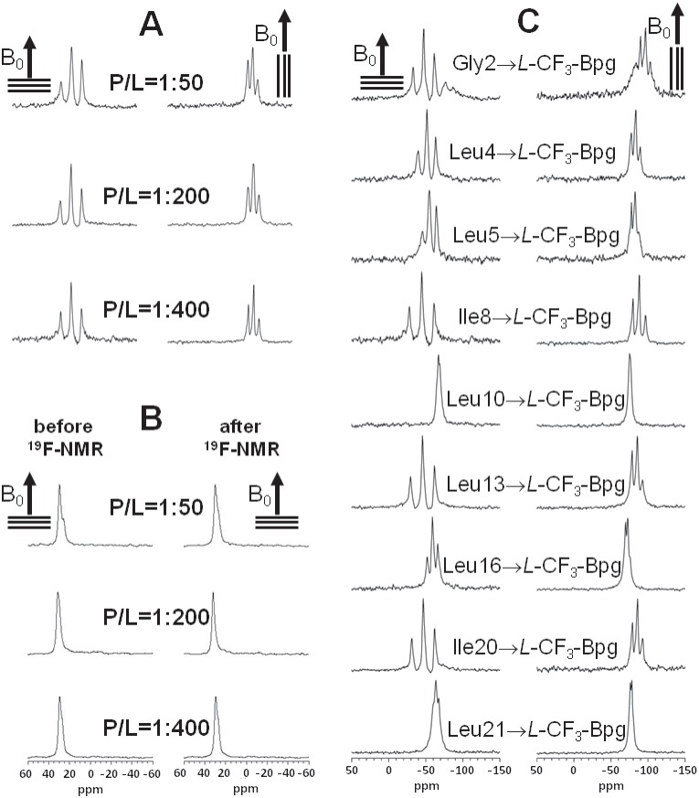
Figure 3. Solid-state NMR spectra of TP10:
(A) 19F-NMR spectra of TP10 labeled with L -CF3-Bpg at Ile8, recorded at three different peptide-to-lipid molar ratios (P/L = 1∶50, 1∶200, and 1∶400) in oriented DMPC/DMPG (3∶1) bilayers. The hydrated membrane samples were aligned with their normal parallel (0°) and perpendicular (90°) to the static magnetic field B0 (indicated by an arrow). (B) Solid-state 31P-NMR spectra of the same samples as in (A), recorded before and after the corresponding 19F-NMR experiment, showing a high quality of lipid alignment. (C) Solid-state 19F-NMR spectra of the nine L -CF3-Bpg labeled TP10 analogs at P/L = 1∶400, from which the dipolar couplings of the CF3-groups were obtained for the structure calculation. All experiments were performed at 40°C.

