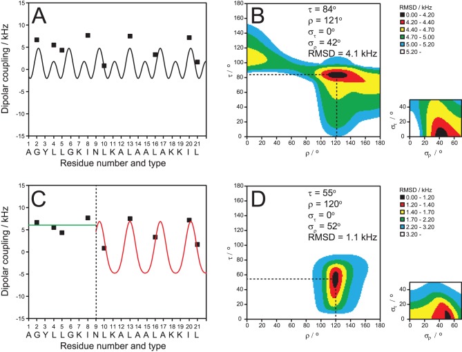
Figure 4. NMR structure analysis of TP10.
Dipolar wave analysis of the 19F-NMR dipolar couplings of monomeric TP10 in hydrated membranes (DMPC/DMPG, P/L = 1∶400). It is not possible to fit all nine L -CF3-Bpg labels to a continuous α-helix (A), as a poor fit with a high RMSD would be obtained (B). A fit of the five C-terminal labels (C, red) produces a good result with a low RMSD (different color scale compared to B) (D). The alignment of the C-terminal α-helix is described by a tilt angle τ≈55° and an azimuthal rotation angle ρ≈120°, with a moderate wobble (σρ) around the long axis. The black region of the plot shows the possible range of τ and ρ with the same RMSD. The N-terminal region of TP10 is intrinsically unstructured in the plane of the membrane, as seen from the characteristic uniform dipolar splittings of around +7 kHz (C, green).

