
Figure 5. Membrane-bound structure of TP10, as derived by solid-state 19F.
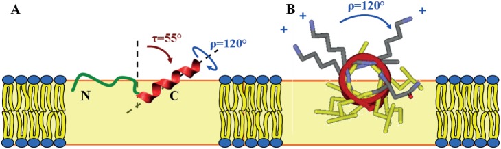
-NMR. (A) The N-terminal region is intrinsically unstructured (green) and connected to the C-terminal α-helix (red). The amphiphilic helix is embedded in the lipid membrane with a tilt angle of τ≈55° and an azimuthal rotation angle of ρ≈120°. (B) The helical wheel projection of the C-terminal mastoparan part illustrates how the charged Lys residues (grey) point towards the aqueous phase, while the hydrophobic residues (yellow) face the interior of the membrane. The yellow box represents the bilayer (not drawn to scale, and without implying any particular insertion depth of the peptide within the bilayer).