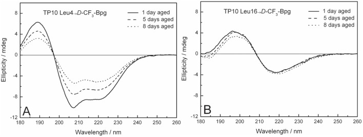
Figure 7. OCD spectra of TP10.

Representative OCD spectra of TP10 labeled with D -CF3-Bpg in oriented DMPC/DMPG (3∶1) bilayers at P/L = 1∶50, measured after 1, 5, and 8 days of ageing. (A) Peptide analogs with a substitution in the N-terminal region (here: position Leu4) have a predominantly α-helical structure, just like the WT peptide. (B) When D -CF3-Bpg is placed into the C-terminal region (here: position Leu16), the peptide aggregates with a β-sheet conformation typical of amyloid-like fibrils.
