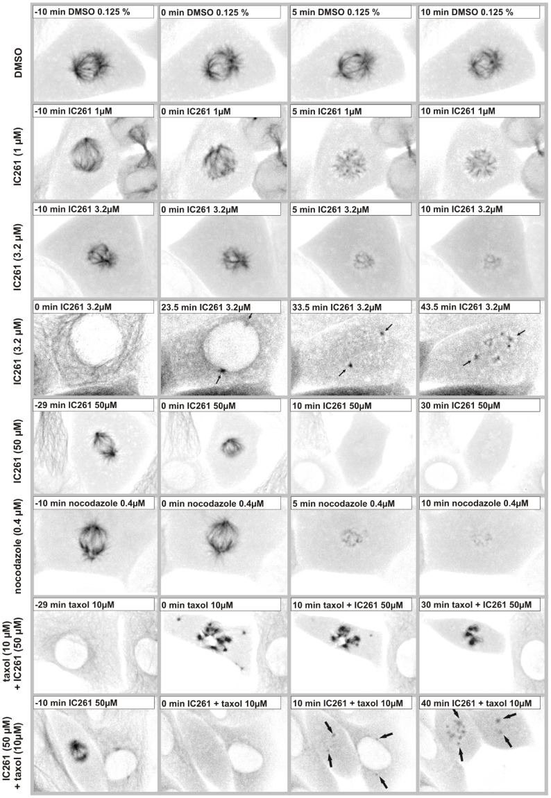
Figure 4. Microtubule destabilizing effect of IC261 in mitotic cells.
CV-1 cells expressing EYFP-tubulin were cultured in a flow-through chamber and observed by time-resolved fluorescence microscopy. At time point “0 min” cells were treated with DMSO (0.125%), IC261 (1 µM, 3.2 µM, 50 µM), 10 µM taxol or 0.4 µM nocodazole. Here exemplary cells are shown for indicated time points (see video sequence, movies S3 and S4). Treatment with low concentrations of IC261 induced a depolymerization of spindle microtubules within a few minutes (row 2–3) in a concentration dependent manner and interestingly by nocodazole treatment a similar phenotype could be observed (row 6). Cells entering mitosis during IC261 treatment had spindle poles and microtubule nucleating centers, but could not form a spindle (row 4, arrows indicate spindle poles). Treatment with 50 µM IC261 induced the complete depolymerization of microtubules within a few minutes (3–5 min, row 5). When cells were treated with 10 µM taxol during time period “−10 min” to “0 min” prior to treatment with taxol+IC261 at time point “0 min” the MT depolymerizing effect of IC261 could be blocked (row 7). When cells were first treated for 10 min with IC261 resulting in a complete dissolution of the spindle apparatus, and subsequently treated with taxol+IC261 tubulin could re-polymerize at the spindle poles (arrows) and in other MT nucleation centers within the cell (row 8).

