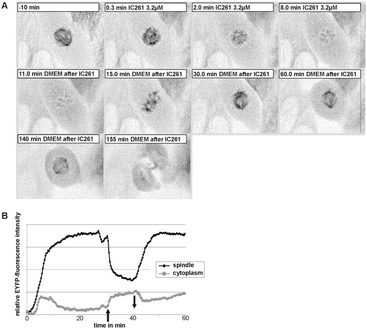
Figure 5. Microtubule depolymerization by IC261 treatment is reversible.
(A) CV-1 cells expressing EYFP-tubulin were treated at time point “0 min” with 3.2 µM IC261 and observed by time-resolved fluorescence microscopy (see video sequence, movie S5). The spindle apparatus of the representative cell shown here was dissolved within 8 min. At time point “10 min” IC261 was removed by exchange of media. Within a few minutes spindle MTs were built up again (“15 min”) and 20 min after removal a morphologically unimpaired spindle apparatus had been developed (“30 min”). After 2 h the cell proceeded into anaphase and cytokinesis (“155 min”). (B) Densitometric analysis of grey values. For quantitative analysis the relative mean intensity of EYFP-tubulin fluorescence signal in a defined region of interest (ROI) around the spindle apparatus and in the cytoplasm was measured by the software CellR. Due to IC261 treatment at time point “0 min” (arrow up) the relative intensity immediately decreased due to MT depolymerization and subsequent removal of IC261 at time point “10 min” (arrow down) lead to a reconstruction of microtubules.

