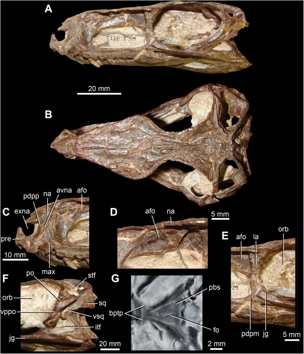
Figure 1.

Anatomy of Gracilisuchus stipanicicorum Romer [8]. A. Skull in right lateral view (reversed). B. Skull in dorsal view. C. Close-up of the right premaxilla and anterior ends of right maxilla and nasal in lateral view (reversed). D. Close-up of the left antorbital fossa above the antorbital fenestra in lateral view. E. Posterodorsal process of the posterior end of the right maxilla in lateral view (reversed). F. Left infratemporal region in lateral view. G. Braincase and posterior end of the palate in ventral view. Abbreviations: afo, antorbital fossa; avna, anteroventral process of the nasal; bptp, basipterygoid process; exna, external naris; itf, infratemporal fenestra; jg, jugal; la, lacrimal; max, maxilla; na, nasal; orb, orbit; pbs, parabasisphenoid; pdpm, posterodorsal process of the posterior end of the maxilla; pdpp, posterodorsal process of the premaxilla; po, postorbital; pre, premaxilla; stf, supratemporal fenestra; sq, squamosal; vppo, ventral process of the postorbital; vsq, ventral process of the squamosal. A-F. MCZ 4117. G. cast of PULR 08.
