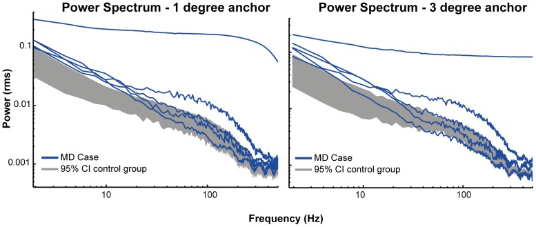
Figure 7. Power spectral density plots.
Power spectral densities for the one and three degrees condition. The grey bars represent the 95% confidence interval (CI) of the control group mean. Each blue line represents one JMD patient. This figure shows that JMD patients had significant more power in the lower frequencies, indicating that low frequency eye-movements dominate the differences in displacement and fixation stability.

