
Figure 6. Microinjection of rAAV-GFP-Cre in the CeA does not affect dendritic spine morphology.
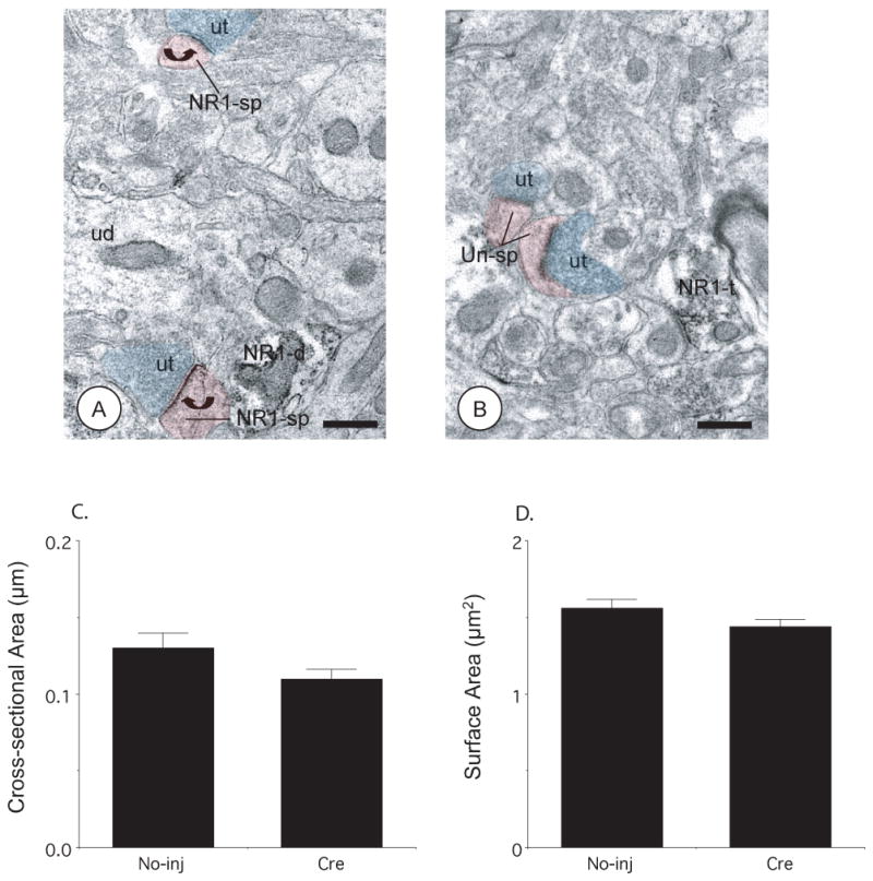
(A–B). Dendritic spines in the uninjected and rAAV-GFP-Cre injected hemispheres show bulbous spine heads, lack intracellular organelles, and receive asymmetric excitatory-type synapse from unlabeled axon terminals (ut, blue shading). The postsynaptic densities of dendritic spines (NR1-sp, red shading) from the CeA not receiving vector microinjection show immunoperoxidase labeling for NR1 (curved arrows), whereas the dendritic spines (Un-sp, red shading) from the contralateral hemisphere receiving rAAV-GFP-Cre do not. Ultrastructural morphometric analysis of dendritic spines showed that there were no significant differences in (C) cross-sectional and (D) surface areas in rAAV-GFP-Cre injected and contralateral hemispheres. NR1-d: NR1 labeled dendritic profile, NR1-t: NR1 labeled axon terminal. Scale Bars= 0.5 μm. Values represent means±SEM