
Figure 2.
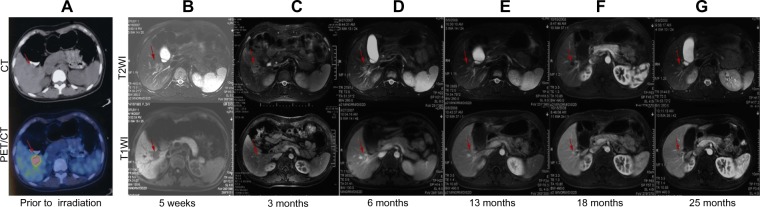
Analysis of initial planning CT and PET/CT as well as follow-up MRI images for a representative patient (a 23-year-old woman treated for a solitary LM from breast carcinoma).
Notes: SBRT was delivered in three fractions of 14 Gy to the 81% isodose line. (A) Initial planning CT and PET/CT images showing a 3.2 cm lesion. (B–G) T1-weighted and T2-weighted MRI images showing a complete response, with decreased attenuation in the surrounding region 6 months after completion of SBRT. The red arrows indicate changes in the liver metastasis after treatment.
Abbreviations: CT, computer tomography; PET/CT, positron emission tomography/computer tomography; MRI, magnetic resonance imaging; LM, liver metastasis; Gy, Gray; PTV, planning target volume; SBRT, stereotactic body radiation therapy.