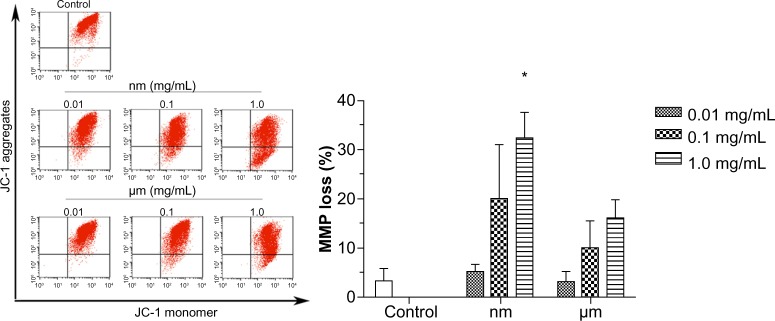
Figure 4.

Effect of SiO2 particles on the mitochondrial membrane potential of BRL cells.
Notes: BRL cells were treated with different concentrations of SiO2 particles for 18 hours, stained with JC-1 and analyzed by flow cytometry. The scatter plot of the flow cytometry analysis shows the distribution of JC-1 aggregates and JC-1 monomer in the mitochondrial membrane and cytoplasm, respectively. The bar graph shows the percentage of JC-1 monomer-positive cells. The results are the mean ± standard deviation of three independent experiments. *P<0.05 versus the control.
Abbreviations: JC-1, 5,50,6,60-tetrachloro-1,10,3,30-tetraethylbenzimidazolecarbocyanine iodide; BRL, buffalo rat liver; MMP, mitochondrial membrane potential.
