
Figure 1.
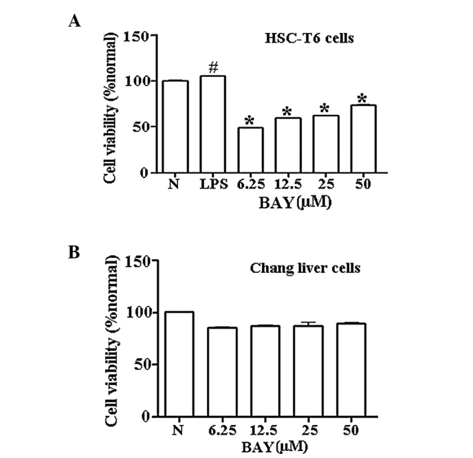
Effect of BAY on cell viability. Cell viability of (A) HSC-T6 cells treated with various concentrations of BAY (6.25–50 μM) after 24-h LPS induction and (B) normal control human Chang liver cells after treatment with BAY for 24 h, as determined by an MTT assay. Data are expressed as mean ± SD. #P<0.01, vs. normal; *P<0.01, vs. LPS-activated HSC-T6 cells. N, normal; LPS, lipopolysaccharide; HSC, hepatic stellate cell; BAY, BAY-11–7082.