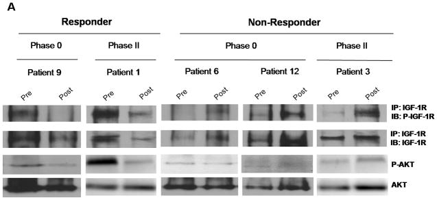
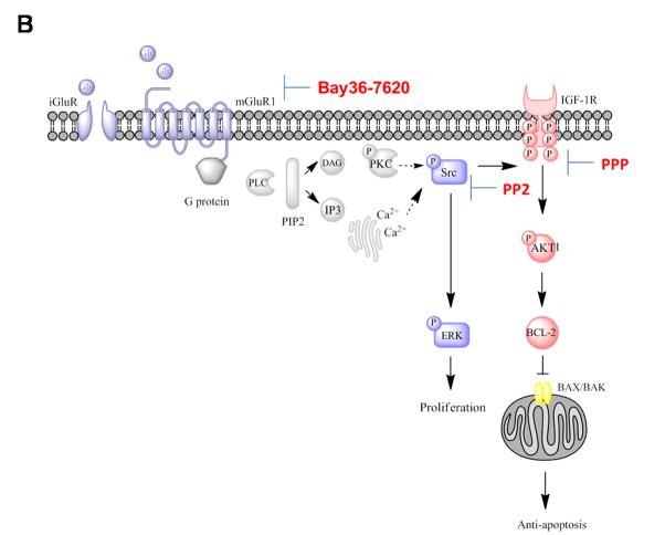
Figure 5. Increased phospho-IGF-1R expression in non-responding patients of Phase 0 and Phase II riluzole trials.

(A) Paired tumor samples from pre-treatment and post-riluzole treatment were analyzed for P-IGF-1R or P-AKT expression by Western immunoblots. A reduction in levels of P-IGF-1R and P-AKT was detected in Phase 0 responder (Patient 9) and Phase II patient with stable disease (Patient 1). In contrast, non-responders in Phase 0 (Patient 6 and 12) and Phase II (Patient 3) riluzole trials showed elevated P-IGF-1R and P-AKT levels or unaltered P-AKT in Patient 6. The blots were subsequently stripped and probed with IGF-1R and AKT antibodies as control. (B) Proposed signaling pathways activated by mGluR1 and mediated by IGF-1R transactivation (Adapted from review article(Teh and Chen, 2012)).

