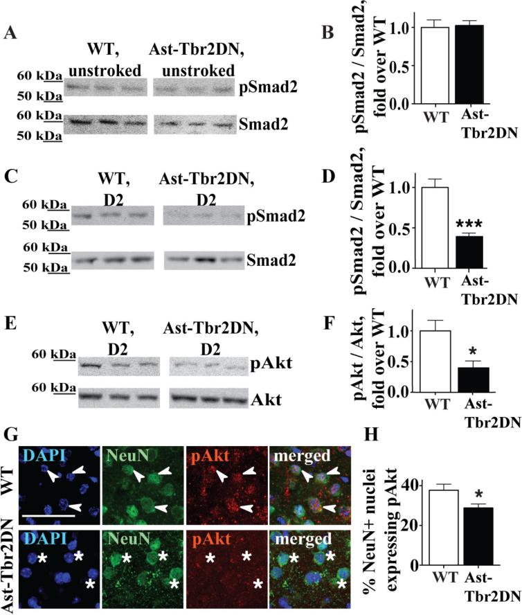
Figure 5. Inhibiting astrocytic TGFβ signaling reduces global TGFβ signaling in the peri-infarct cortex.
A-D. Quantification of the TGFβ downstream signaling mediator Smad2 and its activated phosphorylated form, pSmad2, in the peri-infarct cortex of wildtype controls and Ast-Tbr2DN mice at baseline (A, B) and 2 days after dMCAO (C, D). Representative Western blot images (A, C) and quantification (B, D). E, F. Representative Western blot images (E) and quantification (F) of the TGFβ downstream signaling mediator Akt phosphorylation in the peri-infarct cortex of wildtype controls and Ast-Tbr2DN mice 2 days after dMCAO. G. Representative images of pAkt co-localization with the neuronal nuclear marker NeuN and nuclear marker DAPI in wildtype controls and Ast-Tbr2DN mice 2 days after dMCAO. Scale bar, 20 μm. Arrows, wildtype neuronal nuclei showing pAkt immunostaining. Asterisks, Ast-Tbr2DN neuronal nuclei showing no pAkt immunostaining. H. Quantification of pAkt co-localization with neuronal nuclear marker NeuN in wildtype controls and Ast-Tbr2DN mice 2 days after dMCAO. N=5–6 mice per group. Bars, mean ± SEM. *P<0.05, ***P<0.001, Student's t-test.

