Figure 3.
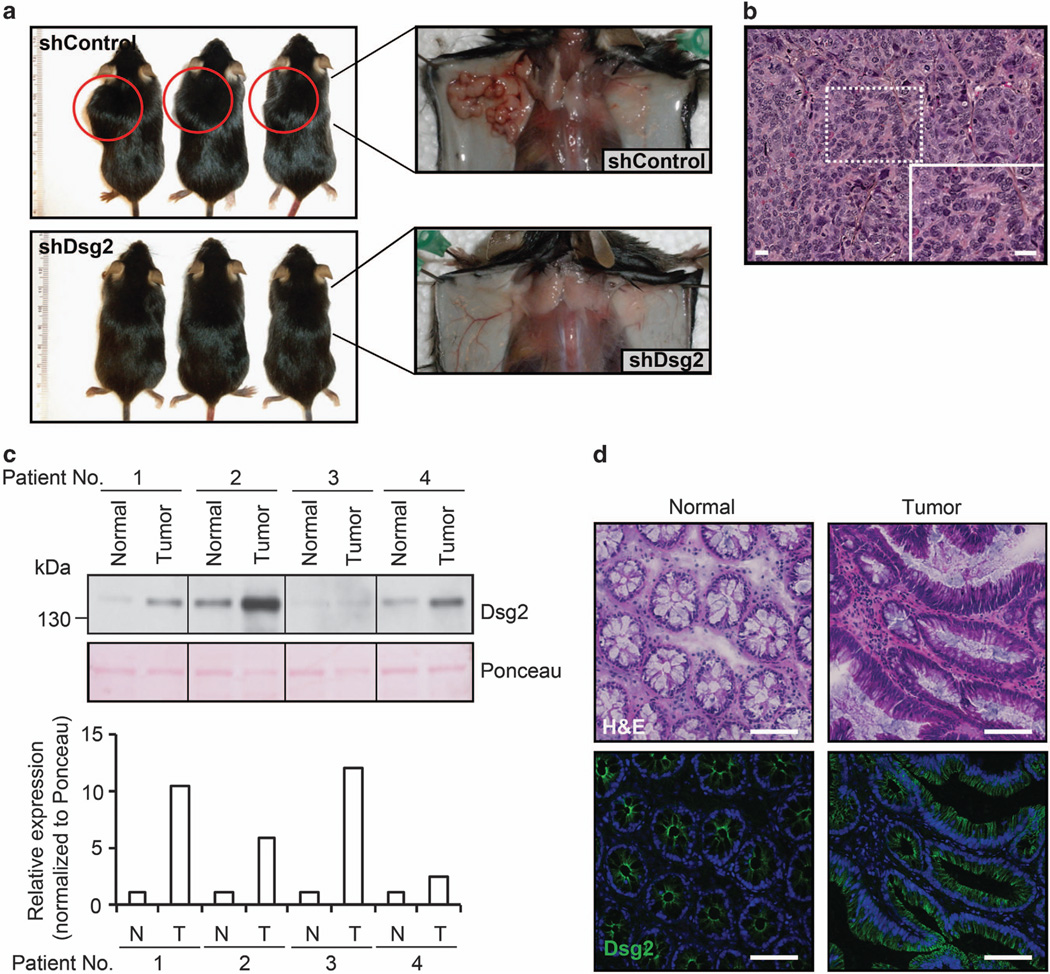
Dsg2-deficient tumorgenic SW480 colon cancer cells fail to grow as tumors in vivo. (a) In vivo xenograft tumor growth for shControl versus shDsg2 cells in Rag1−/− mice. Eight-week-old male mice were injected subcutaneously with 1 × 10⁶ shControl or shDsg2 SW480 cells. Animals were killed at 21 days post injection and the extent of tumor development was assessed macroscopically. All animal experiments were performed in accordance with protocols approved by the Emory University IACUC. (b) Histological analysis of shControl-derived tumors stained with hematoxylin and eosin (Sigma-Aldrich). The insert is a high magnification image of the tumor. Scale bar is 20 µm. (c) Immunoblot analysis of Dsg2 protein in human colonic adenocarcinoma tumors and corresponding normal colonic tissue samples (Protein Biotechnologies Inc., Ramona, CA, USA). The samples were subjected to immunoblot analysis against Dsg2 clone DG3.10 (PROGEN Biotechnik, Heidelberg, Germany). Ponceau staining (Sigma-Aldrich) was used to show equal loading of protein. The histogram shows the relative Dsg2 expression normalized to Ponceau as determined by densitometric analysis. N, normal; T, tumor. (d) Immunofluorescence labeling to localize Dsg2 in normal human colon and human colonic adenocarcinomas (Tumor). Nuclei were stained using To-Pro-3 iodide (TOPRO, Invitrogen). Green, Dsg2 (AH12.2); blue, TOPRO. Consecutive sections of human colonic adenocarcinoma and normal colonic tissue were stained with hematoxylin and eosin. Scale bar is 100 µm.

