
Figure 3.
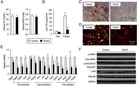
Liver-specific FIP200 deficiency does not exacerbate diet-induced hepatic steatosis. A, Plasma triglyceride and NEFA concentrations under fasted condition. B, Liver triglyceride content of HFD-fed control (white columns) and FILKO (black columns) mice under fed and fasted conditions. C, Oil Red O staining of liver sections from control and FILKO mice under fasted condition. D, SRS microscopy imaging of liver from control and FILKO mice under fasted condition. Lipid droplets were shown as yellow dots in hepatocytes. E, qPCR analysis of mRNA expression of genes involved in lipid metabolism in control and FILKO liver under fasted condition. Data in A, B, and E represent mean ± SE. *, P < .05. F, Immunoblots of total lysates from control and FILKO mouse livers under fed condition. TG, triglyceride.